Can You Have Gout With a Normal Uric Acid Test?
A joint can be hot, swollen, and intensely painful even when the blood urate result is not elevated. Urate may be lower during an acute flare, so a single “normal” result does not by itself exclude gout. The timing of the test and the clinical findings matter.[1][2]
A high result alone also cannot explain every painful joint. The aim is to choose tests that answer the remaining clinical questions, not simply collect more scans. Possible joint infection requires prompt assessment, separate from any plan to repeat a blood test later.[1][3]
1. Why might urate be lower during a flare?
A blood test measures the concentration present when the sample is collected. It does not directly measure all the urate crystals already deposited in tissues or identify the cause of today’s painful joint. Because urate can fall during an attack, clinicians interpret the result alongside the history and examination.[1][2]
Bring the date symptoms began and the date blood was taken. Explain whether you were already using urate-lowering treatment. Avoid treating a laboratory reference range, a diagnostic clue, and a gout treatment target as interchangeable. Instead ask, “How does this result change the likelihood of gout, and what remains uncertain?” A lower result than last year does not answer that question on its own.[1][5]
2. When might a repeat blood test help?
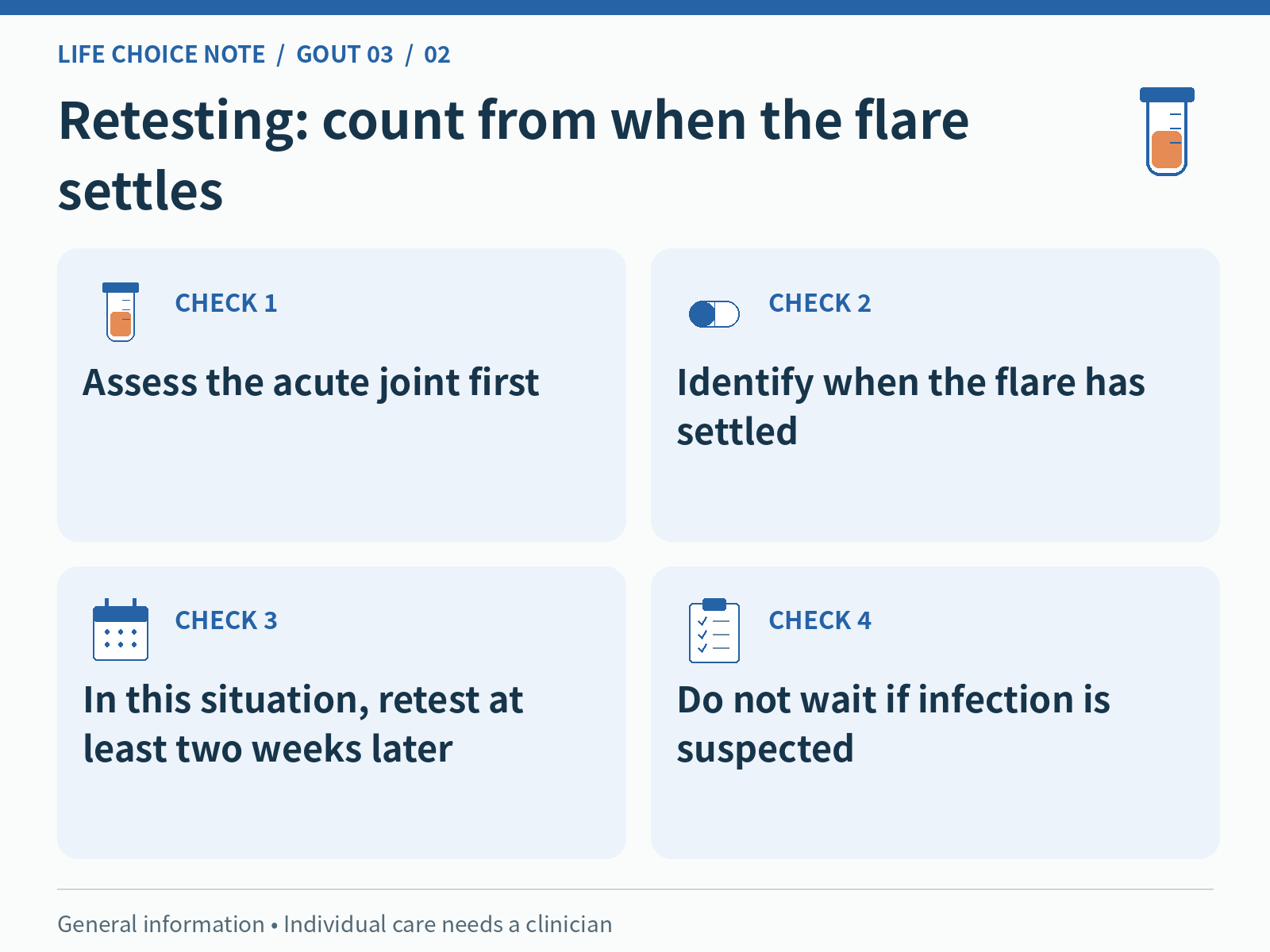
UK NICE guideline NG219 recommends repeating serum urate at least two weeks after a flare has settled when the level during the flare is below 6 mg/dL and gout remains strongly suspected. The interval is measured from resolution of the flare, not simply from the first painful day. Agree the actual timing with the clinician.[1]
This is not advice to postpone the first assessment for two weeks. The current painful joint needs evaluation and appropriate treatment now, particularly if infection is possible. If you bring overseas laboratory reports to a clinic in Korea, check the units: 6 mg/dL is approximately 360 micromol/L. Record the unit and date rather than comparing the printed numbers alone.[1][3]
3. What does joint-fluid analysis show?
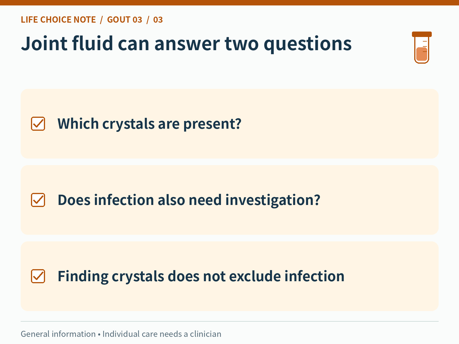
When gout remains uncertain or unconfirmed, a clinician may take a fluid sample from the joint and examine it for crystals. Finding monosodium urate crystals provides important evidence for gout. This is different from repeating a blood urate measurement: the sample comes from the affected joint and can answer additional questions.[1][3][4]
Joint fluid may also be tested for infection when indicated. The presence of urate crystals does not exclude an infection occurring at the same time. Before aspiration, discuss the procedure, expected discomfort, aftercare, allergies, and medicines such as anticoagulants. A small joint or very little fluid can make aspiration difficult, so inability to obtain a sample is not itself a negative gout test.[2][4]
4. How do the imaging options differ?
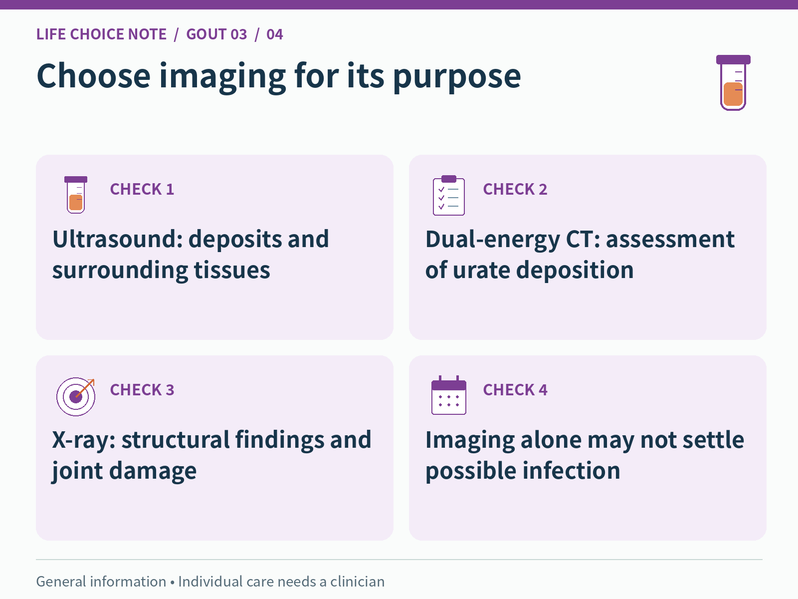
If aspiration cannot be performed or uncertainty remains, imaging may help. Ultrasound assesses deposits and surrounding tissues; dual-energy CT can help identify urate deposition. Plain X-rays can show longer-term joint damage and other structural findings. A normal X-ray early in the disease does not exclude gout. The best test depends on the joint and the clinical question.[1][2][3]
Imaging findings need interpretation rather than a simple pass-or-fail label. EULAR notes that characteristic ultrasound or dual-energy CT findings can support the diagnosis without fluid confirmation in an appropriate setting. It also stresses that aspiration may still be needed to investigate infection or another cause. A scan showing gout does not automatically resolve the question of why a joint is acutely worse today.[3]
5. How should you discuss test selection in Korea?
The most expensive or newest test is not automatically the most useful. Ask what uncertainty the proposed test addresses and how positive or negative findings would change the next step. Identifying crystal deposits and excluding an infected joint are related but different goals. Previous records can help the clinician decide what still needs investigation.[1][3][6]
Ask the Korean clinic directly about availability, preparation, expected charges, and insurance coverage; these cannot be guaranteed from a general article. If you had imaging elsewhere, check whether to bring the report and image files. Their purpose is to make the earlier assessment available, not to insist that repeat testing is never necessary. Note the joint and date because two scans with the same name may assess different episodes. If you are unsure whether a previous test confirmed gout or investigated infection, say so explicitly. Ask the clinician to clarify what was established and what remains open.
6. What should you clarify when results return?
Ask whether the conclusion is confirmed gout, probable gout, or a diagnosis that remains uncertain. Find out which results are still pending and how you will receive them. Joint-fluid crystal findings and infection investigations may answer separate questions. Improvement in pain is welcome, but it should not become a reason to miss an agreed result review.[3][4]
Leave with a clear next step: when to repeat urate if appropriate, whether another assessment is needed, and which changes should trigger earlier contact. New fever, shivering, feeling worse, or rapidly worsening joint pain requires prompt reassessment rather than waiting for a booked scan. The safest interpretation combines the results with your current condition and an explicit follow-up plan.[1][3]
What to bring to your appointment
- Dates of symptom onset, improvement, and blood sampling
- Urate value, units, and laboratory reference range
- Current medicines and previous gout investigations
- The specific question each test is intended to answer
- Pending results and how to obtain them
- Repeat-test timing and reasons for earlier reassessment
Common questions
Does a urate result below 6 mg/dL during a flare rule out gout?
No. If suspicion remains strong, NICE recommends repeating it at least two weeks after the flare settles. Immediate assessment of the joint, including possible infection, should not be delayed.[1]
If urate crystals are found, can infection be ruled out?
No. Gout and joint infection can coexist. Further testing depends on the clinical situation, even when crystals have been identified.[3]
Can a normal X-ray completely exclude gout?
No. X-rays are particularly useful for structural damage. A normal early X-ray does not exclude gout; the need for further testing depends on the whole assessment.[1][2]
This article provides general health information for adults, not an individual diagnosis or prescription. Tests and medicines depend on your symptoms, medical conditions, and other treatments. Seek prompt medical assessment for a rapidly worsening painful joint or possible infection.
Related published reading
- CKD and Supplements: Bring the Bottle, Labs and Medicine List
- Supplements That Can Interfere With Lab Tests: Biotin and Beyond
- Previous: Does High Uric Acid Always Need Gout Medication?
- Next: What Should You Do When a Gout Flare Starts?
Official and primary sources
- Gout: diagnosis and management (NG219), recommendations · NICE · Published 2022-06-09 · Checked 2026-10-15
- Gout: diagnosis and management (NG219), rationale and impact: diagnosis · NICE · Published 2022-06-09 · Checked 2026-10-15
- 2023 EULAR recommendations on imaging in diagnosis and management of crystal-induced arthropathies in clinical practice · EULAR / Annals of the Rheumatic Diseases · Published 2024; recommendations named 2023 · Checked 2026-10-15
- Synovial Fluid Analysis · MedlinePlus, U.S. National Library of Medicine · Last updated 2024-06-03 · Checked 2026-10-15
- Uric Acid Test · MedlinePlus, U.S. National Library of Medicine · Last updated 2026-07-15 · Checked 2026-10-15
- Gout: Diagnosis, Treatment, and Steps to Take · NIAMS, National Institutes of Health · Reviewed 2023-12 · Checked 2026-10-15
- 통풍 · 질병관리청 국가건강정보포털 · Updated 2026-05-18 · Checked 2026-10-15